Most of us gravitate toward hot glue guns in the midst of an intense crafting session, but researchers at Sungkyunkwan University wondered: What if we brought this into the operating room?
Their hypothesis led them to a major discovery; The scientists have developed a modified glue gun that can 3D print bone grafts directly onto fractures during surgery.

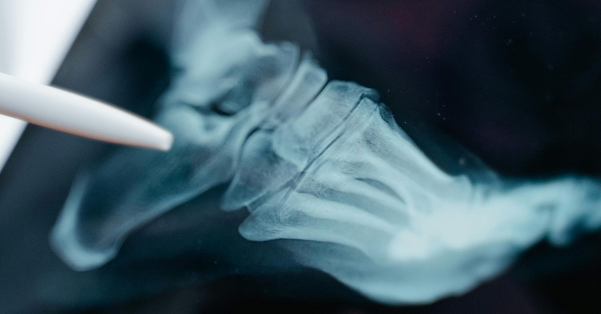
Tested on rabbits, the device was able to quickly create complex bone implants using 3D printing technology, without prefabricating a metal or donor-bone graft in advance.
Placed directly on the area of impact, the 3D-printed grafts offer flexibility while also releasing anti-inflammatory antibiotics and promoting natural bone regrowth at the site, according to the research, published in the journal “Device.”
“Our proposed technology offers a distinct approach by developing an in situ printing system that enables a real-time fabrication and application of a scaffold directly at the surgical site,” Jung Seung Lee, co-author and associate professor of biomedical engineering at Sungkyunkwan University, said in a statement.
“This allows for highly accurate anatomical matching even in irregular or complex defects without the need for preoperative preparation such as imaging, modeling, and trimming processes.”
In other words, even in cases involving irregular bone breaks, the implants from the glue gun can easily fit into place without laborious pre-surgery design and production to ensure a proper fit.
The 3D-printed bone grafts are made from a filament comprised of hydroxyapatite — a feature of the natural bone known to promote healing — as well as polycaprolactone, a “biocompatible thermoplastic” that can liquify in temperatures as low as 60 degrees Celsius (or 140 degrees Fahrenheit).
When applied with the heat-modified gun, this temperature is cool enough to prevent tissue damage during surgery while still being able to conform to the needs of the most complex fractures.

The researchers found the sweet spot. And it speeds up healing and surgical processes, as well.
“Because the device is compact and manually operated, the surgeon can adjust the printing direction, angle, and depth during the procedure in real time,” Lee explained.
“Also, we demonstrated that this process could be completed in a matter of minutes. This highlights a significant advantage in terms of reducing operative time and improving procedural efficiency under real surgical conditions.”
Additionally, using different ratios of the filaments, scientists can customize the hardness and strength of the grafts, meeting different anatomical needs.
Another concern in these kinds of procedures is infection. Therefore, the scientists thought to include two anti-bacterial compounds in the filament: vancomycin and gentamicin.
In their studies, they found that this successfully prevented the growth of E. coli and S. aureus, with the drugs released slowly, allowing them to diffuse into the surgical site over several weeks. That’s right, there were no staph infections reported.
“This localized delivery approach offers meaningful clinical advantages over systemic antibiotic administration by potentially reducing side effects and limiting the development of antibiotic resistance, while still effectively protecting against postoperative infection,” Lee said.
The device was tested on rabbits with severe femoral bone fractures. Over the course of 12 weeks following surgery, the researchers saw no signs of infection and greater bone regeneration than traditional methods.
Of course, for this technology to be adopted clinically for humans, it will require further testing, human trials, and a standardized manufacturing process.
“If these steps are successfully achieved,” Lee said, “we [en]vision this approach becoming a practical and immediate solution for bone repair directly in the operating room.”
You may also like: This student's life-changing injury made him realize how 'outdated' traditional casts are. So he 3D-printed a better one
Header image courtesy of Tima Miroshnichenko/Pexels